
January 29, 2025
By Lynn McCain
When one hears “Artificial Intelligence” – what comes to mind? For some, it may be Terminator-style beings or rogue artificial life forms. In Pathology, however, AI is bringing enhanced patient care and improved satisfaction to pathologists, patients, and clinicians alike. In a recent article in CAP Today, Michigan Medicine’s Department of Pathology was highlighted for its advanced digital pathology workflows. Drs. Ulysses Balis and Mustafa Yousif described the process for initiating digital pathology into our standard of care, which required significant advanced facilities planning, research, development, and investments.
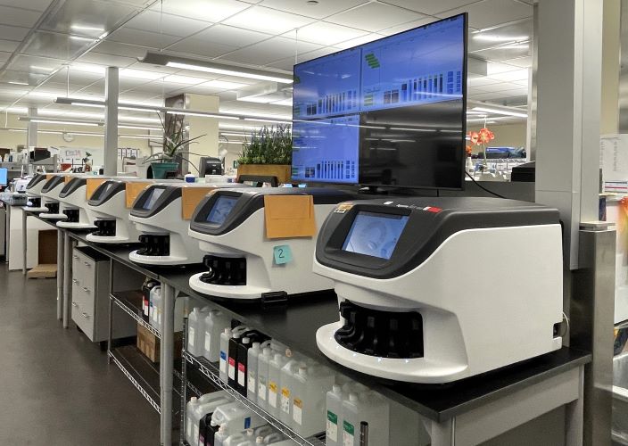
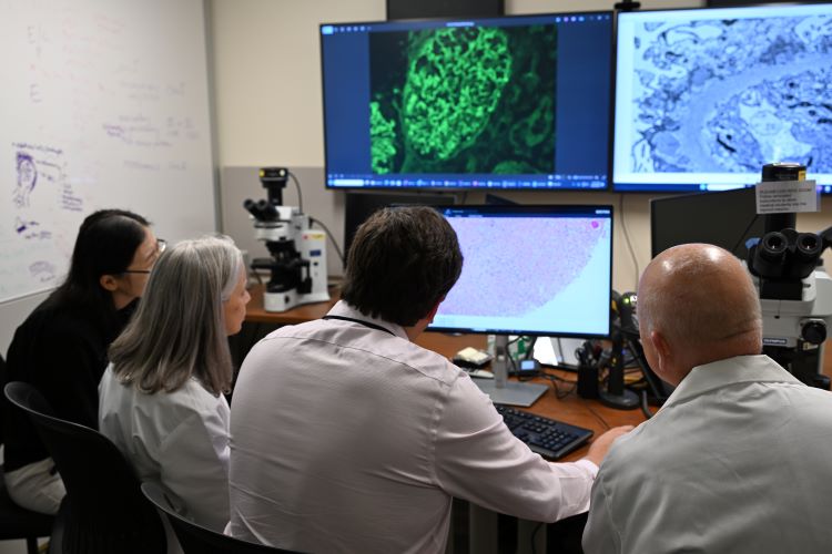
The result is a multimillion-dollar state-of-the-art digital pathology workflow for anatomic pathology cases. This effort leveraged a squadron of digital scanners with a custom flight board embedded within histology, use of standardized DICOM technology to ensure seamless integration, and extensive integration with the laboratory’s existing lab information system to realize seamless integration. The result was the department-wide ability to scan all microscope slides into diagnostic quality digital images. These images are then viewed in spacious sign-out rooms equipped with multiple medical-grade monitors and multi-headed microscopes. When this digital workflow for primary diagnosis is fully deployed across the department and its multiple subspecialty services, the plan is to deploy advanced machine learning / artificial intelligence-driven programs to assist in the QA/QC and diagnostic processes intrinsic to generating anatomic pathology reports. Additional functionality in the image management system allows pathologists with the newfound ability to view multiple stains of the same tissue section simultaneously, thus aiding in the diagnostic process
Additionally, the department’s novel use of the DICOM imaging standard is allowing for pathology images to now be freely sharable across the entire Michigan Medicine health enterprise, thus facilitating patient care needs. In summary, the positive impact of deploying digital pathology for primary diagnosis is being felt not just by pathologists but also by patients and their physicians, who can now more easily communicate about the diagnosis and clearly understand the path ahead.